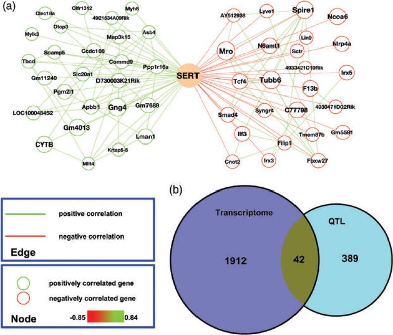
Figure 4. Gene expression networks associated with male midbrain SERT protein expression levels.
(a) Genes (top 25) with highest correlations with SERT protein levels are graphed separately based on the direction of correlation (green: positive; red: negative). Gene node sizes represent the significance of correlation. Gene pairs with correlation coefficients (Spearman rho) above 0.5 are connected by edges (green: positive; red: negative). (b) Venn diagram showing the number of total genes and overlapping genes derived from midbrain SERT gene networks and interval mapping of male midbrain SERT protein levels.

