
Figure 6. Reconstitution of activation of cytosolic PERK protein in vitro.
(A) Recombinant PERK cytoplasmic domain was incubated at a set concentration of IPA. The fold-change in the rate of eIF2α was normalized to the DMSO control and plotted for all concentration. The greatest effects were observed at 500 nM (3.3-fold change) and 3 μM (0.31-fold change) in activity. IPAx showed no effect on the rate of PERK activity at a concentration of 20 μM. (B) Recombinant PERK cytoplasmic domain (2 μM) was preincubated with varying concentrations of IPA (or IPAx) and subjected to chemical cross-linking. An IPA-dependent increase in the dimer, trimer, and tetramer complexes was observed, whereas IPAx (50 μM) showed no effect when compared to the DMSO control.

Figure 6—figure supplement 1. Biochemical reconstitution of PERK activation.
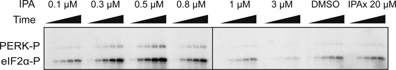
SDS-polyacrylamide gel showing a dose response of IPA. eIF2α phosphorylation was used as a read out of activity. A dramatic activation and inhibition profile is observed. At maximal activity IPA elicits a 3.3-fold change in activation and 0.31-fold change at inhibitory concentrations.
Figure 6—figure supplement 2. The effect of IPAx on PERK activation in vitro.

To test effect of IPAx on PERK activation two concentrations (0.3 and 0.5 μM) were tested in vitro. The SDS-polyacrylamide gel shows the effect of IPAx on PERK activation at 0.3 and 0.5 μM. No activation was observed when normalized to the DMSO control.