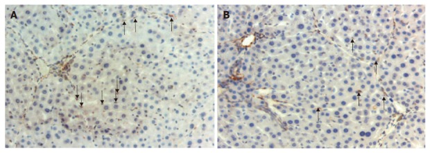
Figure 2.

Activated and apoptotic hepatic stellate cells. A: α-SMA (+) stellate cells in the sinusoids in a section from group 3 (arrows) (× 200); B: Apoptotic activated stellate cells positively stained with TUNEL technique in the sample from group 2 (arrows) (× 200).
