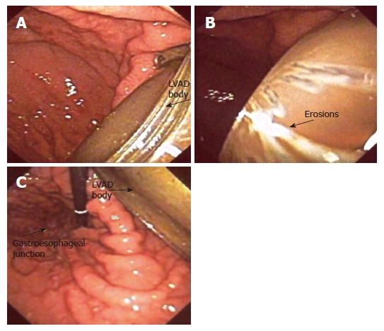
Figure 2.

A: Upper endoscopy study showing the body of the LVAD in the anterior wall of the stomach. The perforation was concealed by the omentum and there were no clinical signs of peritonitis; B: The erosions on the metallic body of the device suggested long exposure to gastric acidic content; C: Endoscope retroflexion revealed that the LVAD had entered the stomach cavity anteriorly and close to the gastroesophageal junction.
