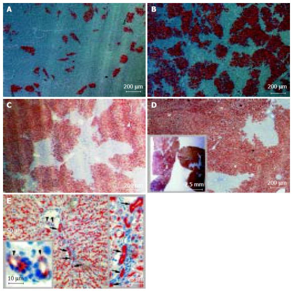
Figure 1.

Long-term liver repopulation 1-20 mo after hepatocellular Tx and hepatic epithelial reconstitution from donor hepatocytes. Recipient DPPIV-deficient Fischer rats were treated with retrorsine and subjected to two-thirds PH. DPPIV-positive donor hepatocytes were infused through the portal vein. 2?06 donor cells per recipient in A-C and 1?06 in D and E. Cryostat sections show DPPIV-positive cells as red-brown after histochemical staining. Data presented as mean± SD from three to six individuals in two or three independent experiments. Tx: transplantation; DPPIV: dipeptidyl peptidase IV. A: 1-mo repopulation: 14.8?.2%; polarization contrast image; B: 2-mo repopulation: 50.7?3.9%; polarization contrast image; C: 3-mo repopulation: 70.4?.0% with hematoxylin counterstaining; D: 20-mo repopulation: 89.8?.7% with hematoxylin counterstaining. The inset box displays the cross-sections of hepatic lobes under stereomicroscopy and E: DPPIV-positive cells not only were expressed in parenchyma but also were found in Hering抯 canal-like structures (arrow) and interlobular bile duct-like structure (arrowhead) in recipient liver; hematoxylin counterstaining.
