
FIGURE 5.
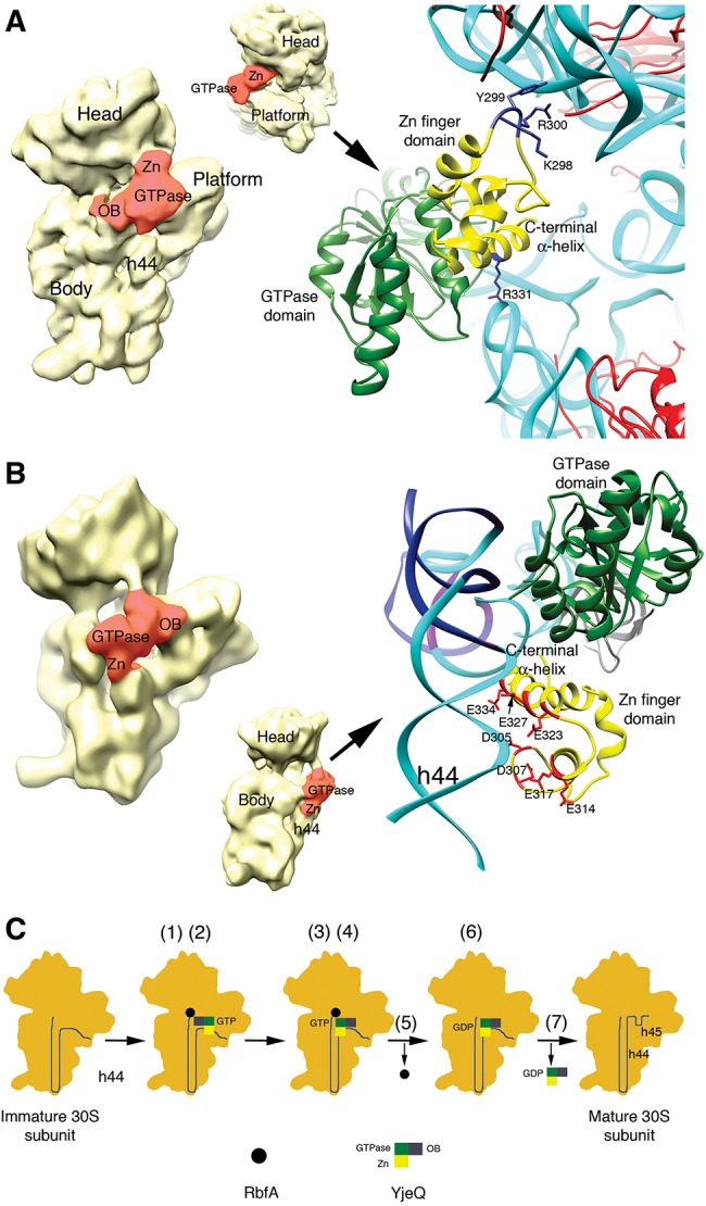
Interaction of the zinc-finger domain of YjeQ with the 30S subunit and working model for the functional interplay between YjeQ and RbfA. Structures of the 30S+YjeQ complex as described in Guo et al. (2011) (A) and Jomaa et al. (2011b) (B). The panel in the left shows a view of the entire complex. Landmarks and domains of the YjeQ protein are labeled. Area colored in red represents the YjeQ protein bound to the 30S subunit. The panel in the right shows a zoomed in view of the complex in the area of interaction of the zinc-finger domain with the 30S subunit. The rRNA is colored in cyan except for some nucleotides (B) that are colored in dark blue. A corresponding density for these nucleotides does not exist in the cryo-EM map of the 30S+YjeQ complex (Jomaa et al. 2011b). The side chains of important residues for this interaction are labeled. Structures shown were obtained from the EMDB (EMD-1884 and EMD-1895) and PDB (2YKR and 4A2I). Images of the structures were prepared with UCSF Chimera software (Pettersen et al. 2004). (C) Proposed model for the functional interplay between YjeQ and RbfA. The model explains the mechanism through which YjeQ facilitates the release of RbfA once the maturation of the 30S subunit is completed. Numbers in brackets refer to the steps of the model described in the text.