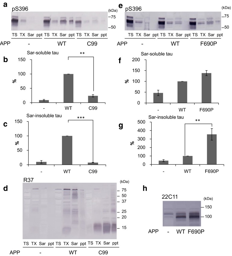
Fig. 6.
Effect of the extracellular domain of APP on intracellular tau aggregation. Immunoblot analysis of lysates from cells transfected with 4R1N tau, cells transfected with both 4R1N tau and APP, and cells transfected with both 4R1N tau and APP-C99. All cells were treated with 4R1N tau fibrils (a). In the lower panel, the Sarkosyl-soluble fraction and Sarkosyl-insoluble fraction detected by pS396 are shown. The results are expressed as means +SE (n = 3). WT-APP was taken as 100 %. **p < 0.01; ***p < 0.001 by Student’s t test against the value of none (b, c). These cells were also detected using R37 (d). Immunoblot analysis of lysates from cells transfected with 4R1N tau, cells transfected with both 4R1N tau and APP, and cells transfected with both 4R1N tau and APP-F690P. All cells were treated with 4R1N tau fibrils (e). In the lower panel, the Sarkosyl-soluble fraction and Sarkosyl-insoluble fraction detected by pS396 are shown. The results are expressed as means +SE (n = 4). WT-APP was taken as 100 %. **p < 0.01 by Student’s t test against the value of none (f, g). The Triton X-100 soluble fractions were detected by 22C11 (h)

