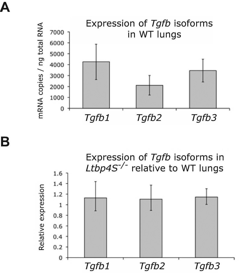
Figure 1.
Expression of Tgfb isoforms in WT and Ltbp4−/− lungs. A. Quantitative RT-PCR for Tgfb isoforms indicated that all three Tgfb isoforms are expressed at similar levels in P0.5 WT lungs. B. The expression levels of Tgfb1, 2 and 3 are similar in both WT and Ltbp4S−/− P0.5 lungs indicating that there is no alteration in Tgfb isoform expression due to the Ltbp-4 loss. Expression levels in B were normalized to levels of G3pdh. Three samples were analyzed per group.

