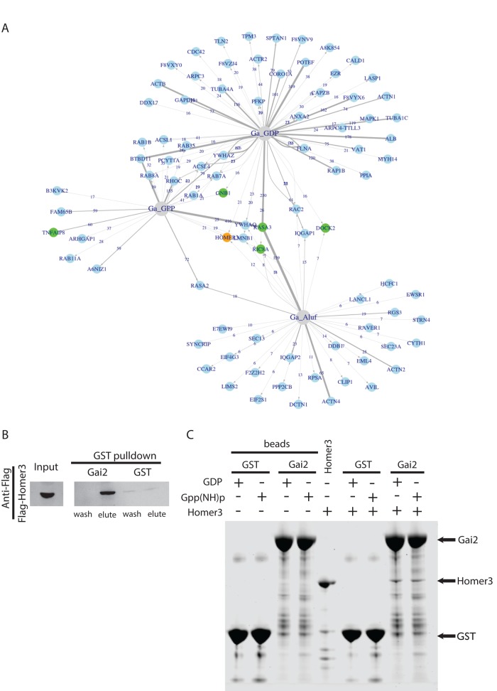
FIGURE 1:
Identification of Homer3 as a neutrophil protein that binds Gαi2. (A) Network analysis of the proteins captured by one or more Gαi2 baits after affinity chromatography in pig leukocyte lysates. The baits are shown in gray, and the proteins are shown in blue. For each protein–bait combination, the numbers at the arrows show the counts, and the width of the lines represents the ZP score. (B) Affinity chromatography with GST-Gαi2 or GST alone for neutrophil lysate containing FLAG-Homer3. Eluted GST-tagged bait plus associated proteins and final wash fraction were subjected to SDS–PAGE and analyzed by immunoblot with anti-FLAG antibody. (C) Affinity chromatography–based test for direct interaction between purified, bacterially expressed Homer3 (prey) and GST-Gαi2 or GST alone (baits). Homer3 directly binds to both GST-Gαi2-GDP and GST-Gαi2-Gpp(NH)p with similar affinity (n = 5; not significantly different). “Beads” refer to baits without Homer3 (prey). Samples were analyzed with SDS–PAGE and stained with CBB. Arrows indicate GST-Gαi2 (66 kDa), Homer3 (47 kDa), and GST (26 kDa).

