FIGURE 6:
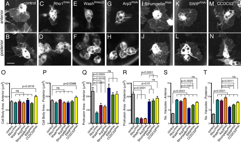
Cellular protrusions are severely reduced in Rho1>Wash>Arp2/3, but not SHRC, posterior hemocytes. (A–N) Confocal projections of anterior (A, C, E, G, I, K) or posterior (B, D, F, H, J, L) hemocytes in equivalently staged embryos expressing GFP in control (A, B), Rho1RNAi (C, D), washRNAi(2) (E, F), Arp3RNAi (G, H), StrumpellinRNAi (I, J), SWIPRNAi (K, L), and CCDC53RNAi (M, N) backgrounds. Scale bars, 10 μm (A–N). (O–T) Quantification of hemocyte cell body size (O, P), protrusion area (Q, R), and number of vacuoles (S, T) in anterior (O, Q, S) or posterior (P, R, T) hemocytes from control or Rho1RNAi, washRNAi(2), Arp3RNAi, StrumpellinRNAi, SWIPRNAi, and CCDC53RNAi embryos at the onset of posterior to anterior migration. All results are given as means ± SEM; p values are indicated.

