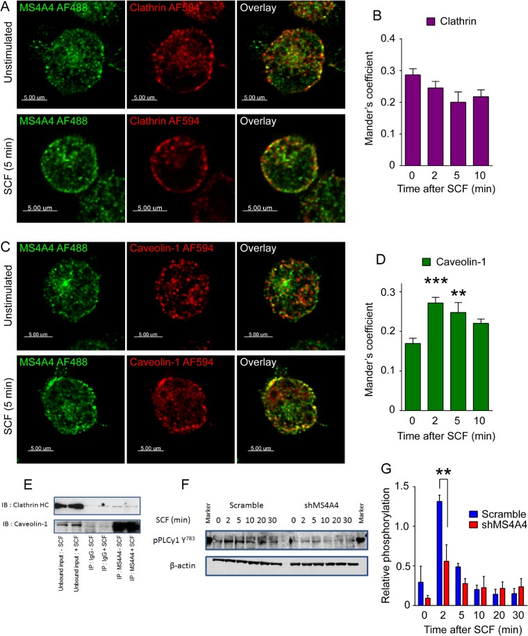
FIGURE 7:
MS4A4 colocalizes preferentially with caveolin-1 over clathrin after stimulation with SCF promoting PLCγ1 phosphorylation. (A) LAD-2 human mast cells immunostained with mouse anti-MS4A4 and rabbit anti-clathrin HC, followed by anti-mouse AF488 and anti-rabbit AF594 before (top) and after SCF stimulation (bottom). No increase in colocalization was observed with stimulation. (B) Manders coefficient of colocalization of MS4A4 and clathrin HC with SCF stimulation time course. (C) LAD-2 human mast cells immunostained with mouse anti-MS4A4 and rabbit anti–caveolin-1 demonstrated an increase in colocalization with SCF stimulation (bottom) compared with untreated cells (top). Scale bars, 5 μm (A, C). (D) Manders coefficient of colocalization of MS4A4 and caveolin-1 with SCF stimulation time course. For B and D, bars are the mean + SEM from the volume of 15 stacks of images from two separate experiments. **p < 0.01, ***p < 0.001, two-way ANOVA. (E) Coimmunoprecipitation using anti-MS4A4 or IgG control as pull down. Immunoblots for clathrin HC and caveolin-1. (F) Immunoblot of PLCγ1 Y783 phosphorylation in response to SCF stimulation over time. β-Actin was used as a control. (G) Densitometry of phosphorylated PLCγ1 (Y783) after correction. Average data from three experiments. Error bars, SEM. **p < 0.01.

