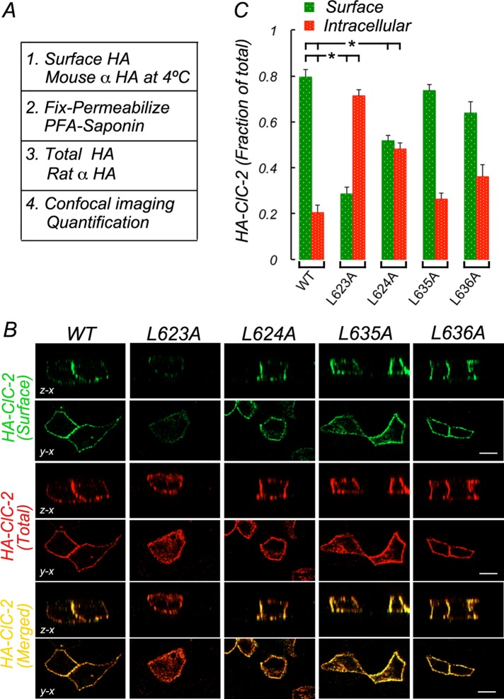
FIGURE 3:
ESMI623LL motif controls basolateral localization of HA-ClC-2. Wild-type and mutant HA-CLC-2 with single leucine residues replaced by alanine in ESMI623LL or QVVA635LL were transiently expressed in MDCK cells allowed to polarize for 4 d in culture. (A) Procedure to sequentially immunolabel HA-ClC-2 before and after saponin permeabilization with mouse and rat antibodies to luminal HA, respectively (see Materials and Methods for details). (B) Surface (green) and total (red) HA-ClC-2 in fully polarized cells. Note that wild-type HA-ClC-2[WT] is restricted to the basolateral membrane, whereas HA-ClC-2 [623L/A] is depleted from the cell surface and accumulated intracellularly. (C) Bars represent fractions of surface and intracellular HA-ClC-2 calculated from confocal images (see Materials and Methods for details). Statistically significant reductions in surface localization of HA-ClC-2 [623L/A] and HA-ClC-2 [624L/A] are indicated. No significant changes in HA-ClC-2 [635L/A] or HA-ClC-2 [636L/A] were observed. Detailed statistical analysis is shown in Supplemental Table S1. Bar, 12 μm.

