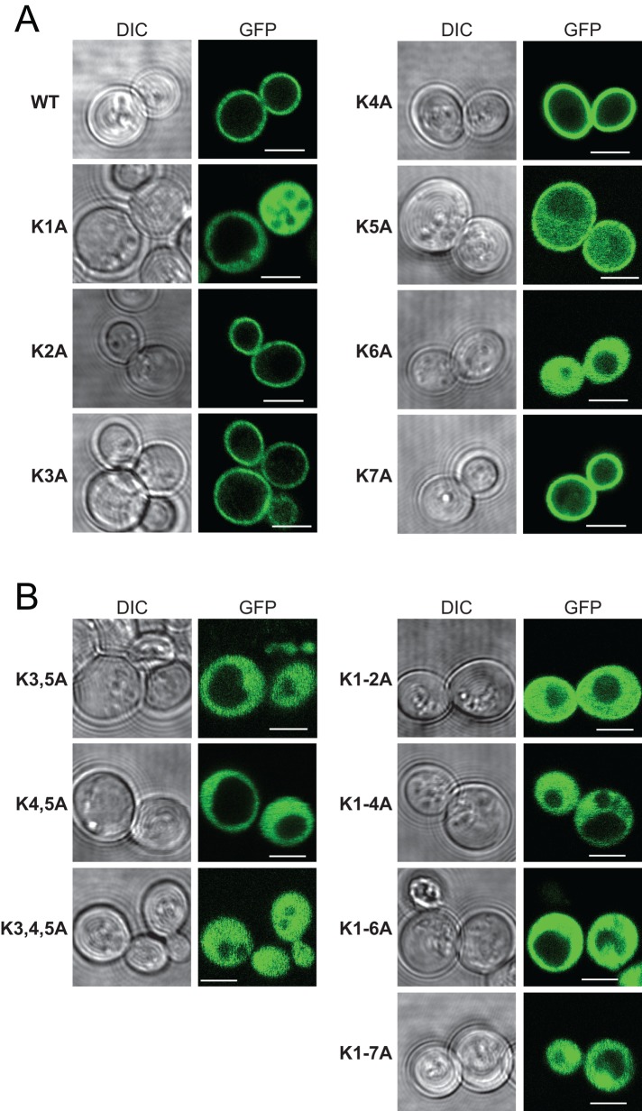
FIGURE 5:
Localization of GFP-tagged WT and the indicated single (A) and multiple (B) K → A substituted nodulin chimeras in WT yeast. Corresponding DIC and GFP confocal image panels. Scale bars, 2 μm. For all mutants, 100–235 cells were imaged and scored. All cells expressing the single K1A and K6A mutant reporters displayed exclusive localization of reporter to the cytoplasm, whereas all cells expressing the K5A reporter showed both PM and cytoplasmic localization for the reporter. Otherwise, >99% of the cells expressing single-mutant K → A derivatives showed exclusively PM localization for the reporter.

