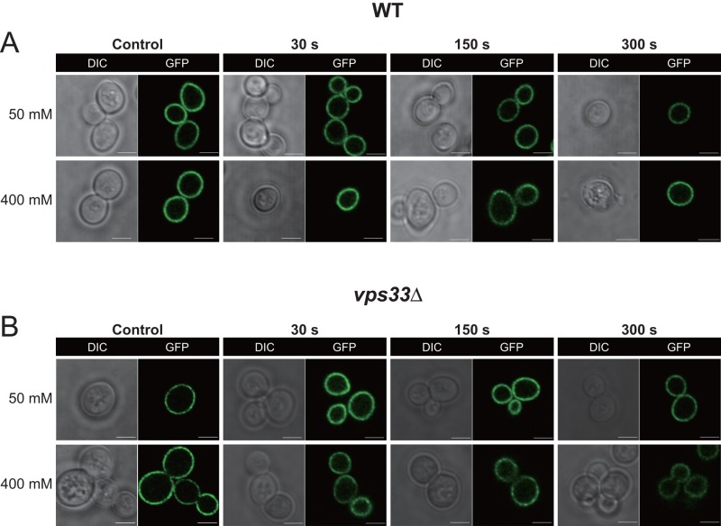
FIGURE 9:
AtSfh1 nodulin domain interactions with phosphoinositide under conditions of Ca2+ influx. Confocal images of WT yeast cells expressing GFP-AtSfh1 nodulin in wild-type (A) and vps33Δ (B) strains, respectively, challenged with 50 or 400 mM CaCl2 as indicated. For both A and B, the control images were taken immediately before Ca2+ challenge. Images were collected every 30 s during a 30- to 300-s post–Ca2+-challenge window of analysis. The time point at which each image was taken is indicated. The vps33Δ mutant strain accumulates high levels of cytosolic Ca2+ under these conditions and sustains these elevated levels throughout the time period the cells were imaged (Miseta et al., 1999). The GFP-AtSfh1 nodulin remained bound to PM in all cells observed for both yeast strains, under both Ca2+ challenge conditions, and at all times imaged. The data are representative of three independent experiments, and 160–326 individual cells were analyzed for each strain, under each condition, for each time point. Scale bar, 2 μm.

