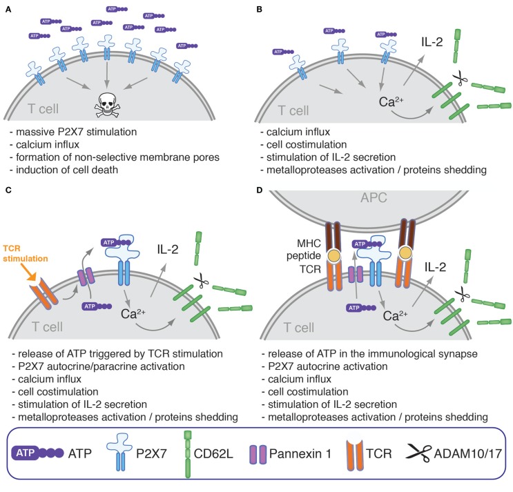
Figure 3.
Modulation of human T cells phenotype, response to mitogenic stimulation, and survival by ATP and P2X7. P2X7-stimulation on the surface of human T cell may have different consequences which could hypothetically depend on the extracellular concentration of ATP, P2X7 density, expression of CD39 and other ATPases, on the nature of T cell subsets, and on their activation status. (A) High concentration of extracellular ATP have been reported to culminate, at least in vitro, in the induction of cell death possibly through the induction of massive membrane depolarization and permeabilization. (B) At lower ATP concentration and/or if cells express a low level of surface P2X7 and/or a high level of ATP-catabolizing enzymes, P2X7-activation could participate instead in T cell costimulation by enhancing the intracellular level of Ca2+, an universal second messenger (76, 78). This may result in the stimulation of NFAT, MAPKs, and IL-2 secretion (69, 70, 72), and in the activation of metalloproteases that catalyze the shedding of CD62L and of other cell surface proteins (79). (C) In some studies, TCR stimulation by mitogenic activators was shown to promote ATP release through pannexin 1 favoring autocrine/paracrine T cell co-stimulation (69, 70, 72). (D) TCR stimulation was also found to stimulate the translocation of pannexin 1 at the immunological synapse together with P2X1 and P2X4 (80). P2X7 is also found within the immunological synapse even if it is not actively translocated within the immunological synapse and remains instead uniformly distributed across the cell surface. This mechanism may thus serve to provide a tonic co-stimulatory signal during physiological activation of a T cell in contact with antigen-presenting cells by promoting the local release of ATP within the immunological synapse, and the local activation of P2X receptors expressed by T cell as well as by antigen-presenting cells.

