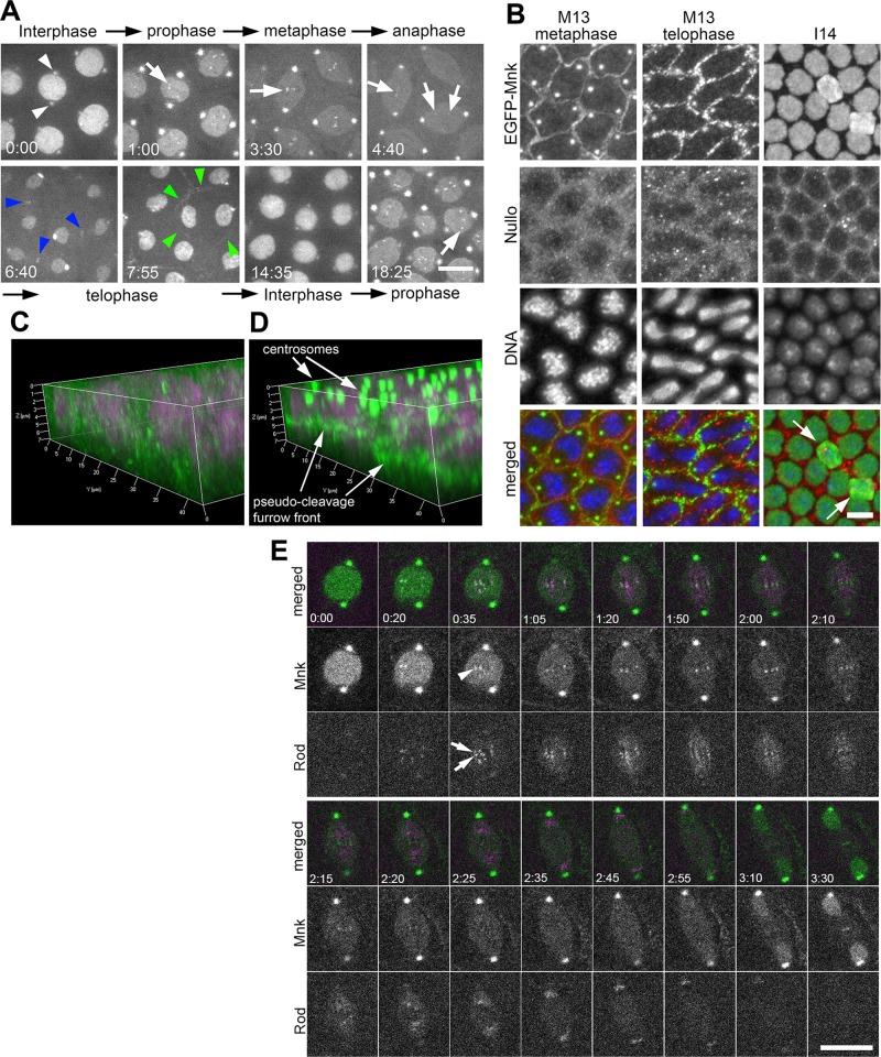
FIGURE 1:
EGFP-Mnk localizes to the nucleus, centrosomes, the interkinetochore/centromere region, the midbody, and pseudocleavage furrows without DNA damage during the syncytial blastoderm stage in Drosophila embryos. (A) Selected frames from a time-lapse spinning-disk confocal movie on a single z-section (Supplemental Movie S1). From cycle 11 interphase to cycle 12 prophase. White arrowheads indicate EGFP signals on centrosomes adjacent to the nucleus during interphase. White arrows indicate EGFP signals on centromeres. Blue arrowheads indicate EGFP signals on the midbody. Green arrowheads indicate EGFP signal on a pseudocleavage furrow surrounding a dividing nucleus. Elapsed time is shown in minutes:seconds. Scale bar, 10 μm. (B) Localization of EGFP-Mnk on pseudocleavage furrows, centrosomes, and interphase nuclei in fixed mnk mutant embryos expressing EGFP-Mnk. Embryos were probed with anti-GFP antibody, anti-Nullo antibody, and DAPI. Each panel presents a maximum projection of z-stacks (M13 metaphase Δz = 7.13 μm; M13 telophase Δz = 5.99 μm; I14 Δz = 8.84 μm). In merges, green indicates anti-EGFP staining, red indicates anti-Nullo staining, and blue indicates DAPI staining. White arrows indicate two nuclei that are dropping from the cortex likely due to spontaneous DNA damage. These nuclei have more EGFP signals than surrounding nuclei (I14). Scale bar, 10 μm. (C) Three-dimensional (3D) reconstruction of Nullo (green) and DNA (magenta) staining. (D) A 3D reconstruction of EGFP-Mnk (green) and DNA (magenta) staining. C and D are reconstructed from the same confocal scanning data shown in B, M13 metaphase. (E) EGFP-Mnk localizes to the interkinetochore/centromere region from prometaphase to anaphase. Frames were selected from a time-lapse confocal movie of an embryo coexpressing EGFP-Mnk and mRFP-Rod (Supplemental Movie S21). White arrows indicate mRFP-Rod that localizes on sister kinetochores, and white arrowhead indicates EGFP-Mnk that localizes between the two sister kinetochores (time, 0:35). Elapsed time is shown in minutes:seconds. Scale bar, 10 μm.

