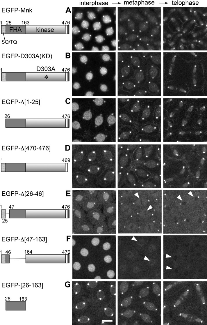
FIGURE 4:
Localization of EGFP-Mnk variants without DNA damage in syncytial blastoderm– stage Drosophila embryos. EGFP was conjugated at the N-terminus of each Mnk variant. Left, schematic diagrams. EGFP was omitted. Selected frames from time-lapse laser-scanning confocal microscope movies that captured EGFP signal during cycle 11 or 12. (A) EGFP-Mnk localizes to the nucleus, centrosomes, interkinetochore/centromeres, the midbody, and pseudocleavage furrows without DNA damage as shown in Figure 1A and Supplemental Movie S1. (B) EGFP-D303A localizes similarly to EGFP-Mnk (Supplemental Movie S7). (C) EGFP-Δ[1-25] localizes similarly to EGFP-Mnk (Supplemental Movie S8). (D) EGFP-Δ[470-476] is not actively transported into the interphase nucleus but localizes to centrosomes, interkinetochore/centromeres, the midbody, and pseudocleavage furrows (Supplemental Movie S9). (E) EGFP-Δ[26-46] localizes to the nucleus, centrosomes, and pseudocleavage furrows but not to interkinetochore/centromeres and the midbody. It is excluded from the area close to mitotic chromosomes (white arrowheads; Supplemental Movie S10). (F) EGFP-Δ[47-163] localizes to the nucleus but not to centrosomes, interkinetochore/centromeres, the midbody, and pseudocleavage furrows. It is excluded from the area close to mitotic chromosomes (white arrowheads; Supplemental Movie S11). (G) EGFP-[26-163] localizes to centrosomes, interkinetochore/centromeres, the midbody, and pseudocleavage furrows but is not actively transported into the nucleus (Supplemental Movie S12). Transgenic proteins were expressed by nos-GAL4VP16 (A) or mTb-GAL4VP16 (B–G) driver in w1118 (A) or mnk mutant (B–G) embryos. Scale bar, 10 μm.

