FIGURE 1:
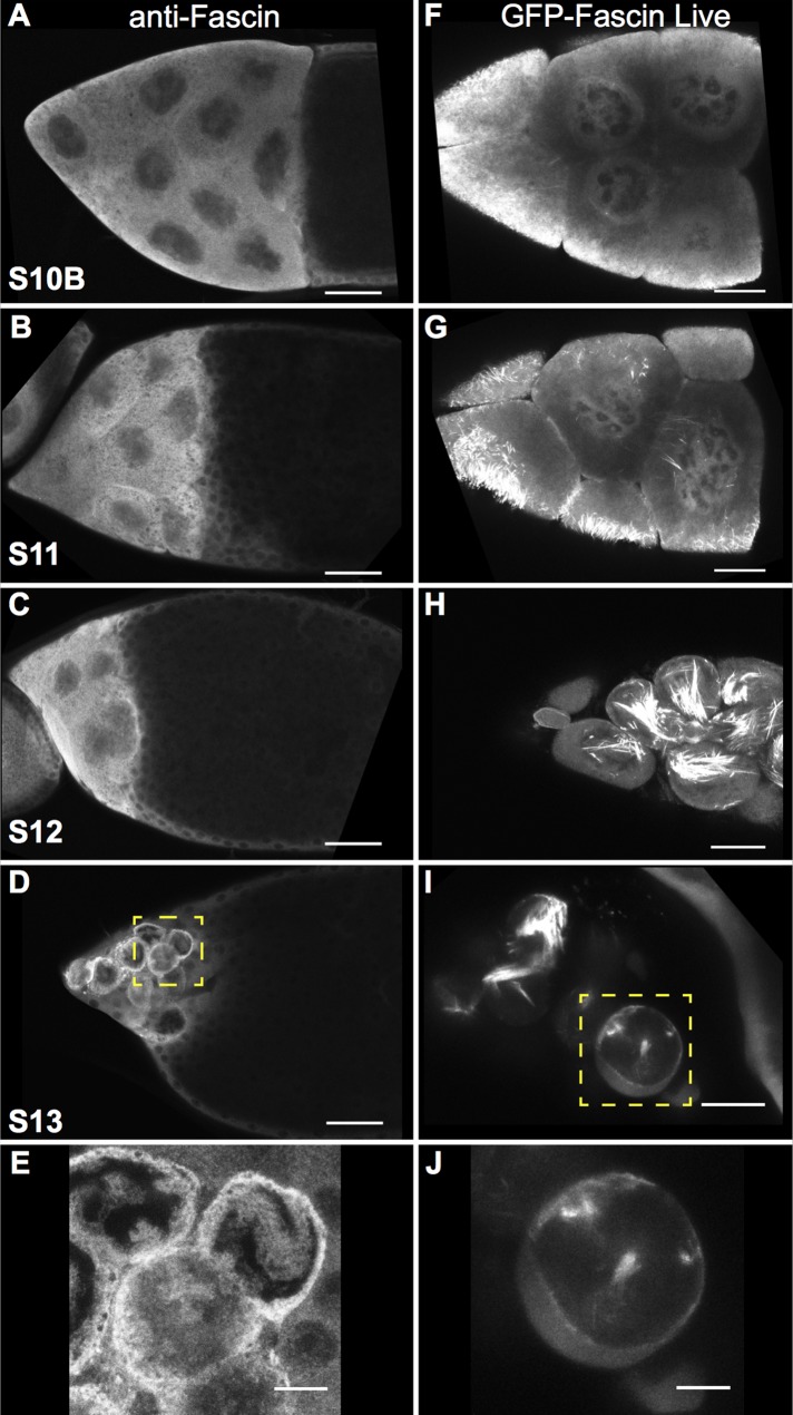
Fascin localizes to the nucleus and nuclear periphery during late-stage follicle development. (A–E) Maximum projections of three to five confocal slices of wild-type late-stage follicles stained with anti-Fascin. (A) S10B, (B) S11, (C) S12, and (D) S13. (E) Zoomed-in image of yellow boxed region in D. Immunofluorescence analysis of Fascin reveals that Fascin localizes not only to the cytoplasm but also to the nucleus during S10B (A) and that nuclear localization increases during S11 and S12 (B, C). At S13, Fascin relocalizes to the nuclear periphery (D, E). Scale bars, 50 μm (A–D), 10 μm (E). (F–J) Maximum projections of three to five confocal slices showing GFP-Fascin (UAS GFP Fascin/oskarGal4) imaged in live follicles using a 40× objective. (F) S10B, (G) S11, (H) S12, and (I) S13. (J) Zoomed-in image of yellow box region in I. Live imaging of late-stage follicles reveals that GFP-Fascin, in addition to being cytoplasmic and on actin bundles, is in the nucleus during S10B–S12 (F–H) and at the nuclear periphery during S13 (I, J). Scale bars, 25 μm (F–I), 10 μm (J).

