FIGURE 7:
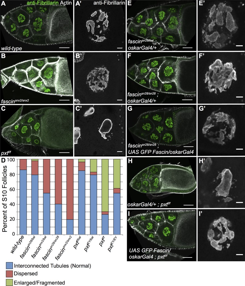
Nucleolar structure is altered in fascin and pxt and mutants. (A–C′, E–I′) Maximum projections of three to five confocal slices at 20× (A–C, E–I) or 63× (A′–C′, E′–I′) of S10B follicles of the indicated genotypes. (A, A′) wild-type, (B, B′) fascinsnx2/snx2, (C,C′) pxtf/f, (E, E′) fascinsn28/bal; oskarGal4/+, (F, F′) fascinsn28/sn28; oskarGal4/+, (G, G′) fascinsn28/sn28; UAS GFP Fascin/oskarGal4, (H, H′) oskarGal4/+; pxtf/f, and (I, I′) UAS GFP Fascin/oskarGal4; pxtf/f. (A–C, E–I) Merged images: Fibrillarin (green) and phalloidin (white). (A′–C′, E′–I ′) Fibrillarin (white). (D) Graph showing quantification of nucleolar phenotype in S10 follicles (blue, normal, interconnected tubules; red, dispersed nucleoli; green, enlarged, fragmented nucleoli). Wild-type nucleoli have an interconnected tubule structure (A, A′), whereas fascin-mutant nucleoli are dispersed (B, B′) and pxt-mutant nucleoli are enlarged and fragmented (C, C′); phenotypic frequencies are quantified in D. Expression of exogenous GFP-Fascin in fascin-null mutants (G, G′) rescues the dispersed nucleolar phenotype (F, F′) and more closely resembles the interconnected tubules most often observed in fascin heterozygous controls (E, E′). Overexpression of GFP-Fascin in a pxt mutant also rescues the enlarged, fragmented nucleoli observed (H, H′ compared with I, I′). Scale bars, 50 μm (A–C, E–I), 10 μm (A′–C′, E′–I′).

