FIGURE 1:
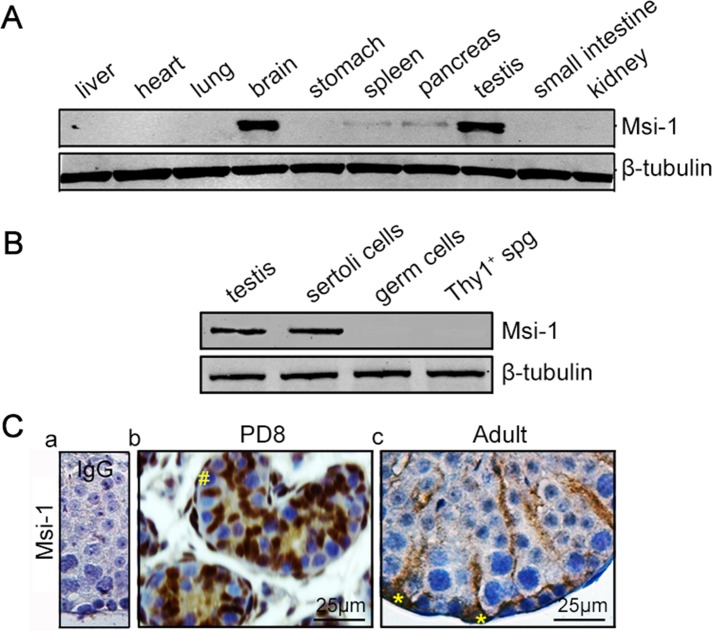
Msi-1 is specifically expressed in Sertoli cells. (A) In mice, Msi-1 was predominantly expressed in the brain and testes, with lower levels detected in the spleen and pancreas. (B) Immunoblotting of Msi-1 in lysates from adult mouse testes, Sertoli cells, germ cells, and Thy1+ spermatogonia; β-tubulin served as a loading control. (C) Immunohistochemical staining of Msi-1 in both immature testes (b) and the seminiferous epithelium of adult mouse testes (c); (a) negative control. The # in b indicates germ cells; * in c indicates Sertoli cell nuclei. spg, spermatogonia; PD, postnatal day. Scale bars: 25 μm.

