FIGURE 5:
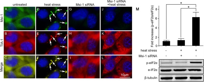
Endogenous Msi-1 protein is essential for stress granule formation. (A–C) In untreated Sertoli cells, Msi-1 and TIA-1 were localized in the cytoplasm. (D–F) Msi-1 was restricted to TIA-1–positive stress granules in the heat-treated Sertoli cells (white arrows). (G–I) Transient transfection of Msi-1 siRNA led to a significant reduction of endogenous Msi-1 levels. (J–L) Stress granule formation was significantly affected in Msi-1–silenced cultures upon heat stress. The nuclei were visualized with DAPI (blue). Scale bars: 10 μm. (M) p-eIF2α was not activated in Msi-1–silenced Sertoli cells exposed to heat treatment. Compared with the group treated with heat alone, p-eIF2α was not activated after Msi-1 knockdown. Histograms summarize the data shown in the panel below after normalizing each data point to β-tubulin. Each bar represents the mean ± SD of n = 3 experiments. *, p < 0.05.

