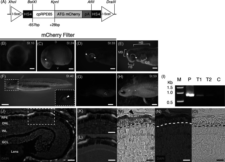
Fig. 3.
RPE65 proximal promoter (−657 bp) assay in F0 transgenic newt larvae (C. pyrrhogaster). a Schematic representation of the transgene construct. b No detection of mCherry expression at stage 10 (blastula). c Stage 24 (early tailbud) onset of mCherry expression localized in the developing optic cup of the dorsal posterior margin, indicated with a white arrowhead. D dorsal, V ventral. d Stage 33 (tailbud) showing mCherry expression in the midbrain and posterior eye margin. e Bright field image of (d). FB forebrain, MB midbrain, HB hindbrain, GS gill slits. (d, e) White arrowheads denote mCherry, and the white dotted arcs indicate the posterior eye margin. f Stage 40 (early swimming larva) showing induction of pigmentation in the eye. Inset (dashed White Square) shows a magnified image of the developing eye expressing mCherry denoted by the white arrowhead (g). h Stage 59 (mature larva) showing a heavily pigmented eye. Yellow arrowhead indicates auto-fluorescence. i PCR detection of cpRPE65-mCherry01 in genomic DNA from stage 59 F0 transgenic larvae. P Positive control cpRPE65-mCherry01 plasmid; T1 transgenic larva genomic DNA sample 1; T2 transgenic larva genomic DNA sample 2; C Negative control wild-type genomic DNA. j Promoter activity in the retinal pigment epithelium (RPE). ONL outer nuclear layer; INL inner nuclear layer; GCL ganglion cell layer. k Magnification of dashed white rectangle in (j). l Magnification of dashed white rectangle in (j), showing mCherry/bright field merged image of RPE apical microvilli. m, n Immunostaining of a retinal section with an anti-mCherry antibody. Signal was visualized by DAB treatment (brown). Black and yellow arrowheads indicate the RPE cell nucleus. o Negative control (without anti-mCherry antibody). Dashed black and white lines in (m, n, o) indicate the apical microvilli of the RPE. Scale bars, b–e 0.5 mm, f 2.0 mm, g–h 0.4 cm, j–o 50 μm. (Color figure online)

