. 2015 Apr;48(4):229–233. doi: 10.5483/BMBRep.2015.48.4.272

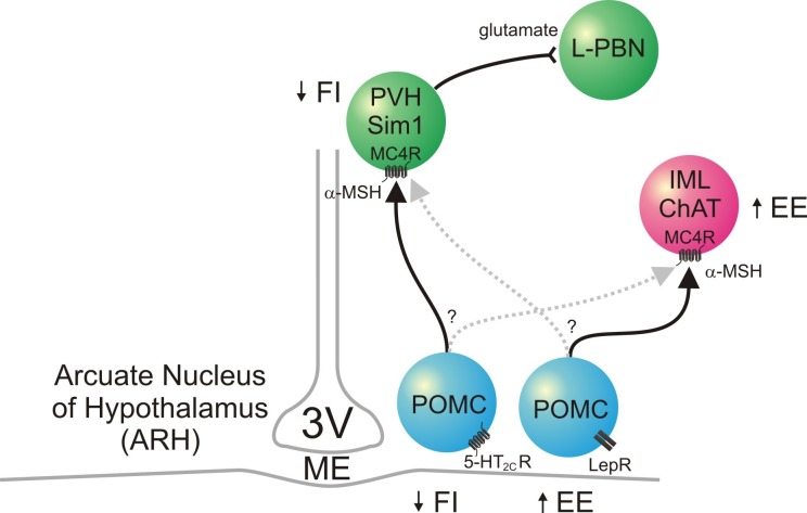
Fig. 1. POMC neurons and the central melanocortin pathway. Two distinct populations of POMC neurons are present within the ARH; 5-HT2CR-expressing POMC neurons suppress food intake, and LepR-expressing POMC neurons increase energy expenditure. The effects of MC4Rs on energy balance are localized to different central nuclei; MC4Rs expressed by the PVH Sim1 neurons suppress food intake, and MC4Rs expressed by the IML sympathetic neurons increase energy expenditure. The solid black lines show possible connections between the specific POMC neurons and specific brain nuclei; the connections represented by the broken gray lines have not been ruled out. MC4R-expressing PVH glutamatergic neurons send axons to the neurons of the L-PBN. 3V: third ventricle, 5-HT2CR: serotonin 2C receptor, α-MSH: α-melanocyte stimulating hormone, ChAT: choline acetyltransferase, EE: energy expenditure, FI: food intake, IML: intermediolateral column of spinal cord, L-PBN: lateral parabrachial nucleus, LepR: leptin receotor, MC4R: melanocortin-4 receptor, ME: median eminence, POMC: pro-opiomelanocortin, PVH: paraventricular nucleus of hypothalamus, Sim1: single-minded 1.

Fig. 1.