
Fig. 3.
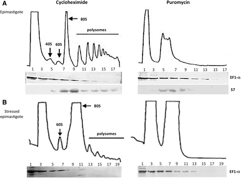
Polysome fractionation. (a) Unstressed epimastigote polysome profile. (b) Stressed epimastigote polysome profile. Fractions were analyzed by western blotting with antiserum against EF-1α (1:300 dilution) and S7 ribosomal protein (1:500). The numbers in the fractions are related to: 1 and 3 – free fraction, 5 – 40 S, 7 – 60 S, 9-17 monosomes to heavy polysomes