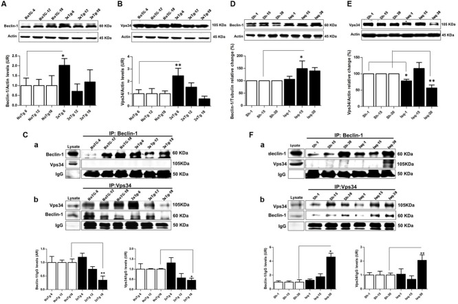
FIGURE 3.
Macroautophagy effects on the cerebral cortex of 3xTgAD mice compared with cerebral ischemia in rats. Representative bands from SS-Cx lysates of 3xTgAD and ischemic rats were evaluated by Western blotting and immunoprecipitation. (A) Beclin-1 (p ≤ 0.043) and (B) Vps34 (p ≤ 0.029) increased in 3xTg-AD at 6 months. (C) The Vps34/Beclin-1 association was lost at 12 and 18 months. (D,E) Beclin-1 increased at 15 days in Ischemic rats (p ≤ 0.029) and Vps34 was reduced at 1 day (p ≤ 0.021) and 30 days (p ≤ 0.007). (F) Association between Beclin-1 and Vps34 was detected at 30 days and Vps34/Beclin-1 complex formation at 15 and 30 days post-ischemia. The data from the 3xTgAD mice and ischemic rats were normalized against the internal control for each time point in the NoTg mice and sham rats, respectively. β-actin was used as the loading control. The data are shown in the bar graph as AUs. IgG was used as the loading control in the immunoprecipitation assay. The data are presented as the mean ± SEM; n = 3–5; ∗p < 0.05; ∗∗p < 0.001.

