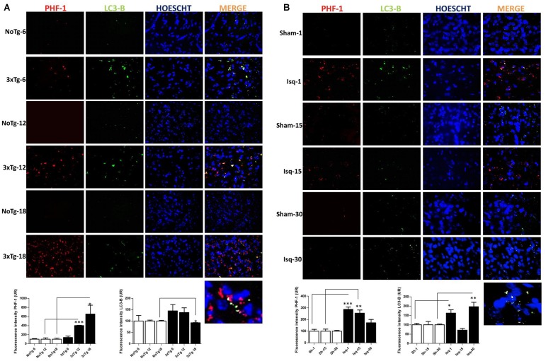
FIGURE 6.
LC3B and PHF-1 immunofluorescence in the cerebral cortex of 3xTgAD mice and in cerebral ischemic rats. LC3B and PHF-1 immunofluorescence was detected in the SS-Cx of 3xTgAD mice and cerebral ischemic rats. (A) PHF-1 fluorescence intensity increased in 3xTg-AD mice at 12 (p ≤ 0.0003) and 18 months (p ≤ 0.0233), and LC3B fluorescence intensity decreased in 3xTg-AD mice at 18 months (p ≤ 0.0499). (B) PHF-1 fluorescence intensity increased at 1 (p ≤ 0.0001) and 15 days post-ischemia (p ≤ 0.0079), and a reduction at 30 days (p ≤ 0.1191) post-ischemia, and LC3B fluorescence intensity increased at 1 day (p ≤ 0.0156) and 30 days post-ischemia (p ≤ 0.0077). The data were compared with the internal control values for each time point in the disease progression. Magnification, 60; scale bar, 20 μm; n = 3. The data were expressed as the mean ± SEM; ∗p < 0.05; ∗∗p < 0.004; ∗∗∗p < 0.001.

