
Figure 1.
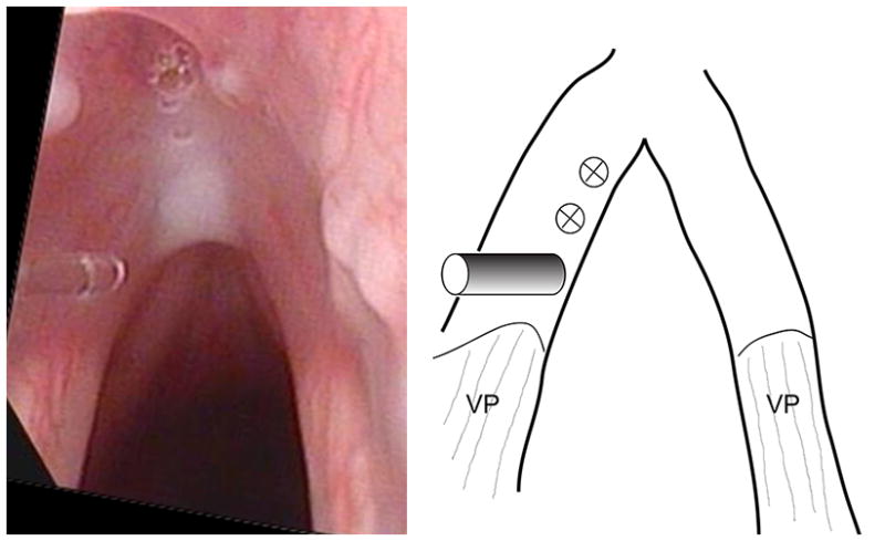
Intraoperative photo of the Ho:YAG fiber in contact with the left vocal fold mucosa (left panel), and schematic showing the locations of laser application (right panel). The fiber is positioned at the most posterior of the 3 locations of laser application, and the two anterior ones are marked in the schematic. VP – vocal process.