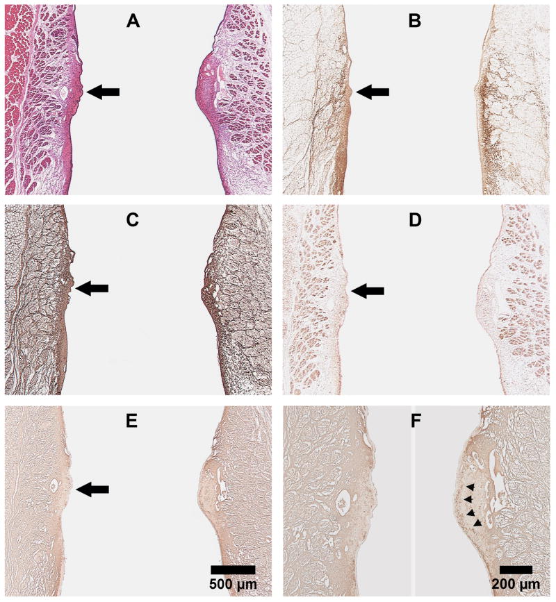
Figure 3.
Representative coronal sections of rabbit larynges harvested 4 weeks after laser treatment. (A) H&E; (B) Collagen type I; (C) Collagen type III; (D) Elastin; and (E) Hyaluronic acid. Vocal fold on left - scarred; right - normal. Panels A-E (25X): Large black arrow points to the location of laser injury. (F) Higher magnification of (E), with arrowheads pointing to the continuous band of HA in the lamina propria of normal vocal fold (100X).

