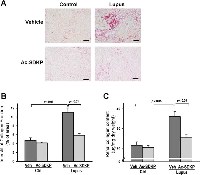
Fig. 6.
Effect of Ac-SDKP on renal collagen. A: representative images of interstitial collagen depositions detected by picrosirius red staining of kidney tissues collected at the end of 20-wk treatment with either vehicle or Ac-SDKP. Shown are digitized sections captured using ×20 microscope objective. Scale bar = 200 μm. B: quantitative analysis of images showed a significantly lower amount of interstitial collagen depositions in Ac-SDKP-Lupus mice, compared with Veh-Lupus mice. Data were calculated as a percentage of the fibrotic area and expressed as means ± SE; n = 6–10. P < 0.01, Veh-Ctrl vs. Veh-Lupus and Ac-SDKP-Lupus vs. Veh-Lupus mice. C: quantitative analysis of total renal collagen content determined by hydroxyproline assay. Significantly lower renal collagen content was observed in Ac-SDKP-Lupus mice compared with Veh-Lupus mice. Data were calculated as a microgram of collagen per milligram of dry tissue weight and expressed as means ± SE; n = 6–10. P < 0.05, Veh-Ctrl vs. Veh-Lupus mice and Ac-SDKP-Lupus vs. Veh-Lupus mice.

