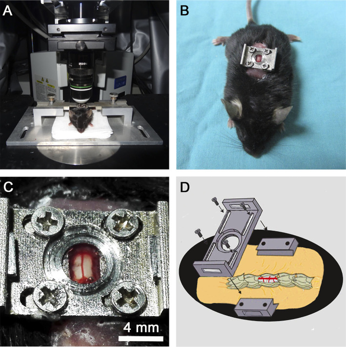
Figure 1. In vivo two-photon imaging of the mouse spinal cord.
(A) The customized spinal stabilization device with an implanted window. (B) The mouse with an implanted window. (C) The segment of the spinal cord exposed for two-photon imaging. (D) The schema showing the implanted window on the exposed T12 spinal cord segment.

