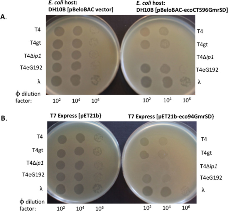
Figure 6. Phage spot tests for T4, T4gt, T4Δip1 (IPI*-deficient), T4 eG192 IPI+ (ΔIPII ΔIPIII), and λvir on E. coli strains expressing EcoCT596GmrSD or Eco94GmrSD.
(A). Two strains DH10B carrying pBeloBAC vector or pBeloBAC-EcoCT596GmrSD were used for comparison. The difference in phage spot (plaques) formation is most evident at 106-fold dilution where EcoCT596GmrSD restricted T4 and T4gt at approximately 5-fold. T4Δip1 (IPI*-deficient) failed to form plaques on EcoCT596GmrSD-expressing strain (input phage ~2–3 × 10⁵ pfu). (B). T7 Express [pET21] and T7 Express [pET21-Eco94GmrSD] strains were used for phage spot tests. Eco94GmrSD moderately restricted T4, T4gt, and T4 eG192, and it did not restrict λvir. T4Δip1 phage was strongly restricted by Eco94GmrSD (no plaque formation at 100-fold dilution, estimated phage input ~2–3 × 10⁵ pfu).

