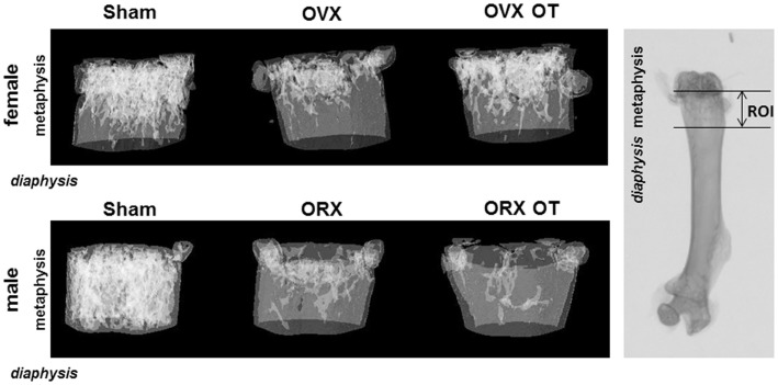
Figure 1.
Micro-computed tomography of distal femur metaphysis of OVX and ORX mice injected or not with OT and their corresponding Sham mice. A three-dimensional representation of a horizontal analysis of femurs from a representative mouse of each group is shown. Right picture displayed a femur radiography with the region of interest (ROI) used for 3D reconstruction.

