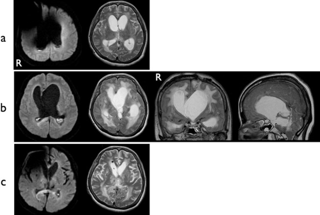
Figure 3.
Case 2. (a) MRI on admission. DWI shows abnormal hyperintensities in the occipital horns of both lateral ventricles. Hydrocephalus is obvious. A metallic artifact is apparent around the right frontal region. (b) Preoperative MRI. Progression of asymmetrical hydrocephalus with findings of ventriculitis is evident. (c) Postoperative MRI. Both ventriculitis and hydrocephalus have resolved. DWI, diffusion-weighted imaging.

