
Figure 1. Impact of Cell Density on TGF-β Signaling.
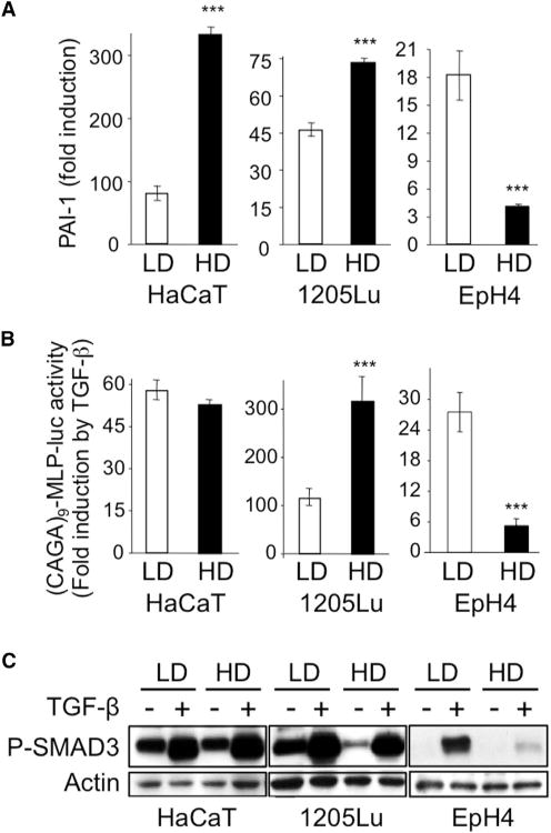
HaCaT keratinocytes, 1205Lu melanoma cells, and EpH4 mouse mammary epithelial cells were grown in either low (LD) or high (HD) density conditions prior to TGF-β (5 ng/ml) stimulation.
(A) Quantitative RT-PCR analysis of PAI-1 expression after a 24-hr TGF-β treatment. Results are expressed as -fold induction by TGF-β in each culture condition and are the mean ± SD from three independent experiments, each measured in triplicate.
(B) Effect of TGF-β on SMAD3/4-specific transcription. Results are expressed as -fold activation of transiently transfected (CAGA)9-MLP-luc activity 18 hr after TGF-β addition to the cultures. Results are the mean ± SD of two independent experiments, each performed with triplicate samples.
(C) Western analysis of P-SMAD3 levels without or with 30 min TGF-β stimulation. Actin levels were measured as a control for the specificity of P-SMAD3 changes under each experimental condition. Results from one representative of several independent experiments are shown.