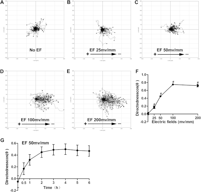
Figure 1. The characteristics of keratinocyte galvanotaxis.
Keratinocytes were stimulated with or without direct current EFs (25, 50, 100 or 200 mV/mm) for 6 h (G) or 3 h (others). (A–E) The migration trajectories of keratinocytes guided by EF (B–E) or not (A), with the starting points positioned at the origin. (F) Quantitative analysis of directedness (cosθ) of keratinocyte migration. (G) The time course of keratinocyte directedness. The data are from at least 100 cells in 3 independent experiments and are shown as the mean ± SEM. #, p < 0.01 compared with the cells without EF stimulation.

