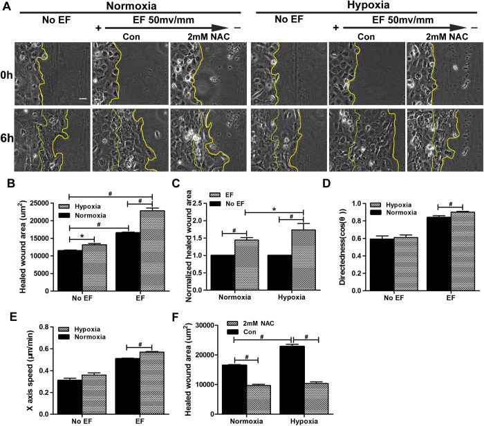
Figure 5. Hypoxic preconditioning accelerates EF-guided keratinocyte migration in monolayer wounds.
Monolayer keratinocytes preconditioned by hypoxia (2% O2, 6 hours) or not were scratch-wounded with a P200 Gilson pipette tip and then exposed or not to an EF of 50 mV/mm for another 6 hours in the absence or presence of NAC. (A) Typical experimental images showing wound edges immediately following and 6 hours post-wounding. The wound edges are illustrated with a yellow line. (B) Analysis of healed wound areas. (C) Analysis of normalized healed wound areas. (D–E) Analysis of the directedness and X-axis speed of keratinocyte migration. (F) Analysis of the healed areas in the absence or presence of NAC. The data are from at least 3 independent experiments and are shown as the mean ± SEM. #, p < 0.01. Bar = 25 μm.

