
Fig. 4.
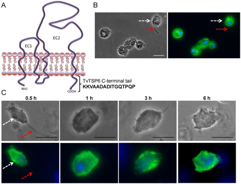
Subcellular localization of TvTSP6ΔCt in absence and presence of host cells using immunofluorescence microscopy.
A. The predicted structure of TvTSP6 in the plasma membrane is shown. EC1 and EC2 = extracellular domains; cytoplasmic NH2 and C-terminal tails are depicted, and the 16 amino acids deleted from the C-terminal tail of TvTSP6ΔCt are shown.
B. Subcellular localization of TvTSP6ΔCt in the absence of host cells is detected using an anti-HA antibody. Flagella (red arrows) and the main body of the cell (white arrows) were indicated. Note the lack of signal in the flagella.
C. Subcellular localization of TSP6ΔCt in parasites bound to VECs. No change in localization is observed. One hundred parasites were counted in triplicate in four independent experiments. Scale bar, 10 μm.