
Fig. 5.
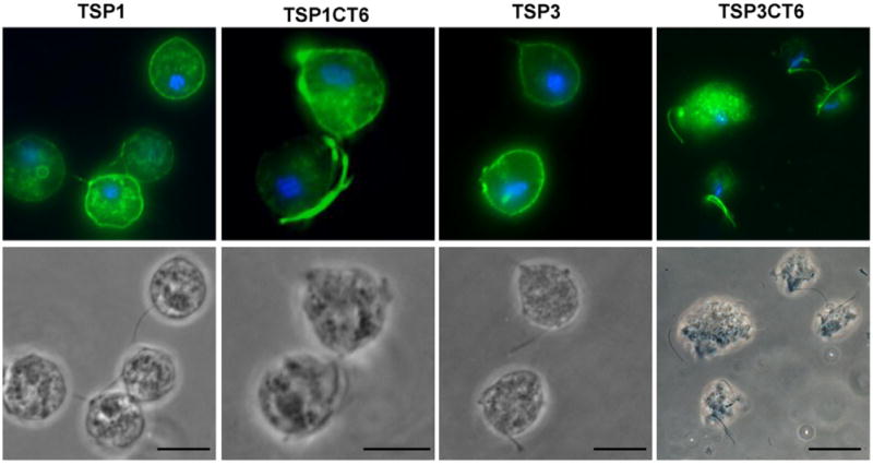
Subcellular localization of TSP1, TSP1CT6, TSP3 and TSP3CT6 in absence of host cells. Cells expressing C-terminal HA-tagged versions of TSP3 (TVAG_280860), TSP3CT6 (chimera with the C-terminal tail of TSP3 replaced by the C-terminal tail of TvTSP6), TSP1 (TVAG_019180) and TSP1CT6 (chimera with the C-terminal tail of TSP1 replaced by the C-terminal tail of TvTSP6) were stained for immunofluorescence microscopy using an anti-HA antibody. The nucleus (blue) was also stained with DAPI. Scale bar, 10 μm. Note the intensity of flagella signal on the chimeric proteins TSP1CT6 and TSP3CT6 relative to wild-type TSP1 and TSP3 proteins.