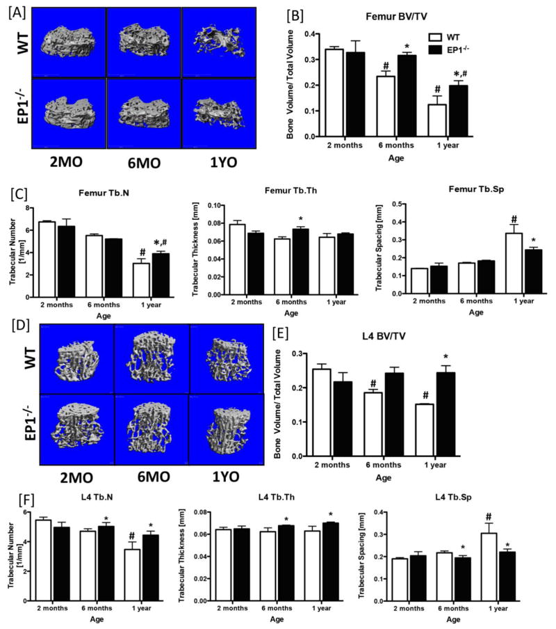
Figure 1. Loss of EP1 is protective against age-induced bone loss.
Three-dimensional microCT reconstructions of the metaphyseal femur [A] and L4 vertebrae [D] of WT and EP1−/− mice at 2-months, 6-months and 1-year of age. [B & E] Bone volume/total volume; [C & F] trabecular number, trabecular spacing, and trabecular thickness quantification of the femur and L4 vertebrae, respectively. N = 5 per group. Statistical analysis was performed using 2-way ANOVA followed by Dunnett’s test. (*) = p < 0.05 v.s. age-matched WT, (#) = p < 0.05 v.s. respective genotype at 2-months.

