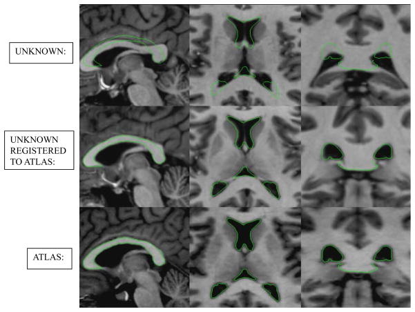
Figure 1. Example of inter-patient image registration. A registration atlas and target temporal bone images are shown, as well as the target image registered to the atlas.
Example of atlas based registration. Shown are delineations of the ventricles in the (left-to-right) sagittal, axial, and coronal views of the MR in the bottom row. These delineations are overlaid on (top row) the MR of another subject and (middle row) the same subject from the top row after registration to the image in the bottom row.

