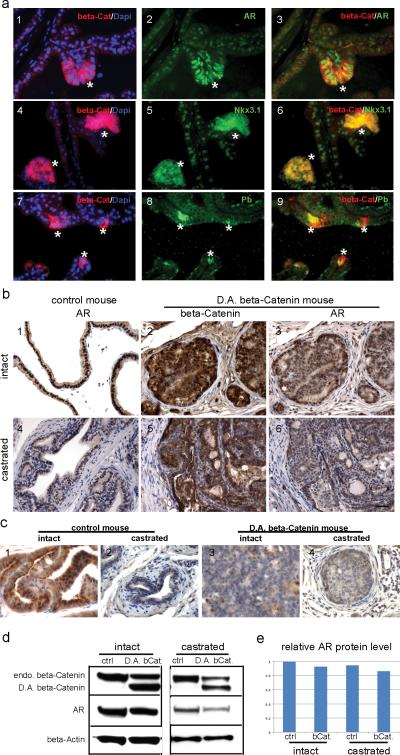
Fig. 5.
AR signaling in PBCre4/ CatnbΔ(ex3) mouse prostate. a, immunofluorescence staining performed on prostate sections from a 12 week old PBCre4/ CatnbΔ(ex3) mouse prostate. 1-3: dual staining of β-Catenin and AR. 4-6: dual staining of β-Catenin and Nkx3.1. 7-9: dual staining of β-Catenin and probasin (Pb). β-Catenin was in red; AR, Nkx3.1 and probasin were in green. At the areas where β-Catenin accumulated in cytoplasm/nucleus (indicated by asterisks), AR was up-regulated compared with other areas that showed only membrane β-Catenin staining. Concurrently, AR target genes-Nkx3.1 and probasin were up-regulated with the activation of β-Catenin. b, immunostaining performed on prostate sections from 22 week old intact (panel 1-3) or castrated (panel 4-6) control and PBCre4/ CatnbΔ(ex3) mice. At this age, AR level was slightly decreased in PBCre4/ CatnbΔ(ex3) mouse prostates compared with control under both intact and castration condition. c, probasin staining. d, western blot to analyze the AR level in intact or castrated control and PBCre4/ CatnbΔ(ex3) mouse prostates. e, quantification of western blot results. Scale bars represent 50 μm.

