
Fig. 1.
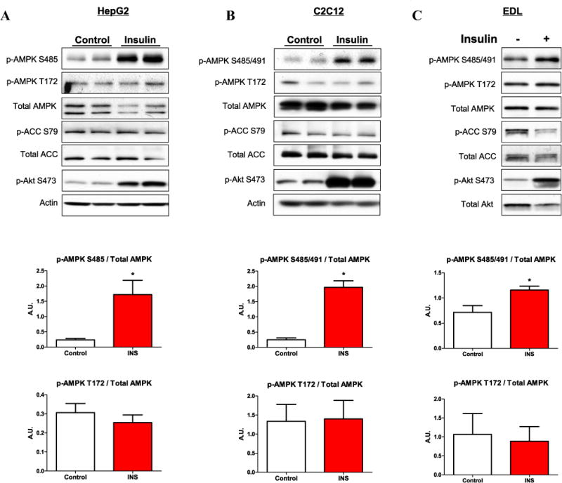
Insulin stimulates phosphorylation of AMPK Ser485 in HepG2 hepatocytes and Ser485/491 in C2C12 myotubes and incubated EDL muscle. HepG2 cells (A) and C2C12 myotubes (B) were cultured in normal glucose (5.5 mM), serum starved overnight, and stimulated with insulin (100 nM) for 15 min. Rat extensor digitorum longus (EDL) muscles were removed and equilibrated in Krebs–Henseleit buffer for 20 min, then stimulated with insulin (10 mU/ml) for 10 min (C). Following cell/tissue lysis, western blot analyses were performed, and representative blots are shown. Densitometry was used to quantify western blots. Phosphorylation of AMPK was normalized to total AMPK, and normalized data are displayed (shown in line with the corresponding cell type/tissue). Results are means ± SE (n = 3–6 per treatment). All experiments were performed in triplicate. *p < 0.05 vs. control.