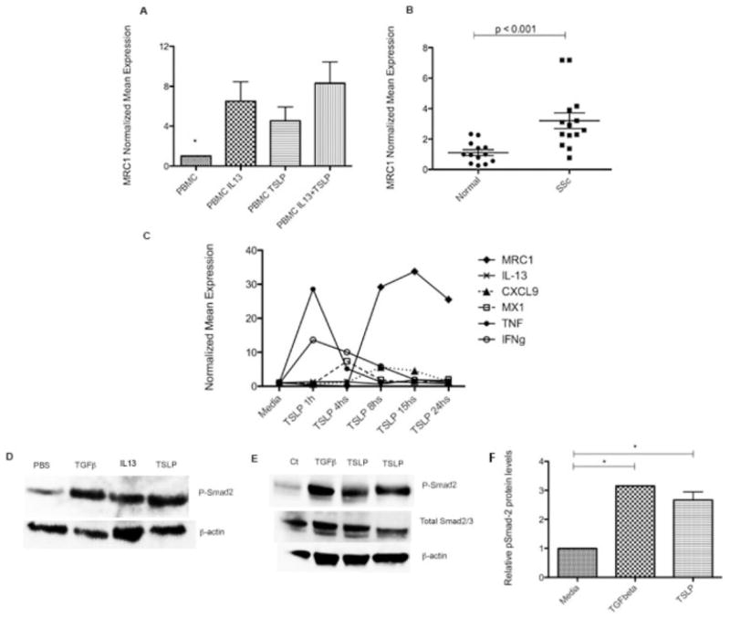
Figure 4.
A, Mannose receptor 1 (MRC1) expression in peripheral blood mononuclear cells (PBMCs) from 5 healthy controls, stimulated for 18 hours with interleukin-13 (IL-13; 20 ng/ml), thymic stromal lymphopoietin (TSLP; 10 ng/ml), or both. Media alone was used as a control. Bars show the mean ± SEM fold change normalized to mRNA expression in controls. * = P < 0.001 versus all other groups. B, MRC1 expression in skin samples from 14 patients with diffuse cutaneous systemic sclerosis (SSc) and 13 healthy controls. Each data point represents a single subject; horizontal lines show the mean and SEM. C, Gene expression in PBMCs from 3 healthy controls, stimulated for 1–24 hours with TSLP (10 ng/ml) using media alone as a control. Values are the mean ± SEM fold change normalized to mRNA expression in the control. TNF β tumor necrosis factor; IFNγ = interferon-γ. D, Higher expression of pSmad2 in the skin of mice subjected to transforming growth factor β (TGFβ), IL-13, and TSLP pump than in mice subjected to phosphate buffered saline (PBS) pump. E, Increased expression of pSmad2 in healthy human dermal fibroblasts stimulated for 1 hour with TGFβ (2.5 ng/ml) or TSLP (10 ng/ml) after overnight starvation at 100% confluence and compared to media alone (Ct). Results are representative of 5 independent experiments. In D and E, blotted proteins were probed with rabbit anti-pSmad2 monoclonal antibody, total Smad2/3 antibody, and secondary antibody, and visualized using enhanced chemiluminescence. As a control for equal protein loading, the membrane was stripped and reprobed for β-actin using a monoclonal antibody for β-actin. F, Expression of pSmad2, quantified by scanning densitometry and corrected for levels of β-actin in the same samples of human dermal fibroblasts. Bars show the mean ± SEM. * = P < 0.05.

