Figure 6.
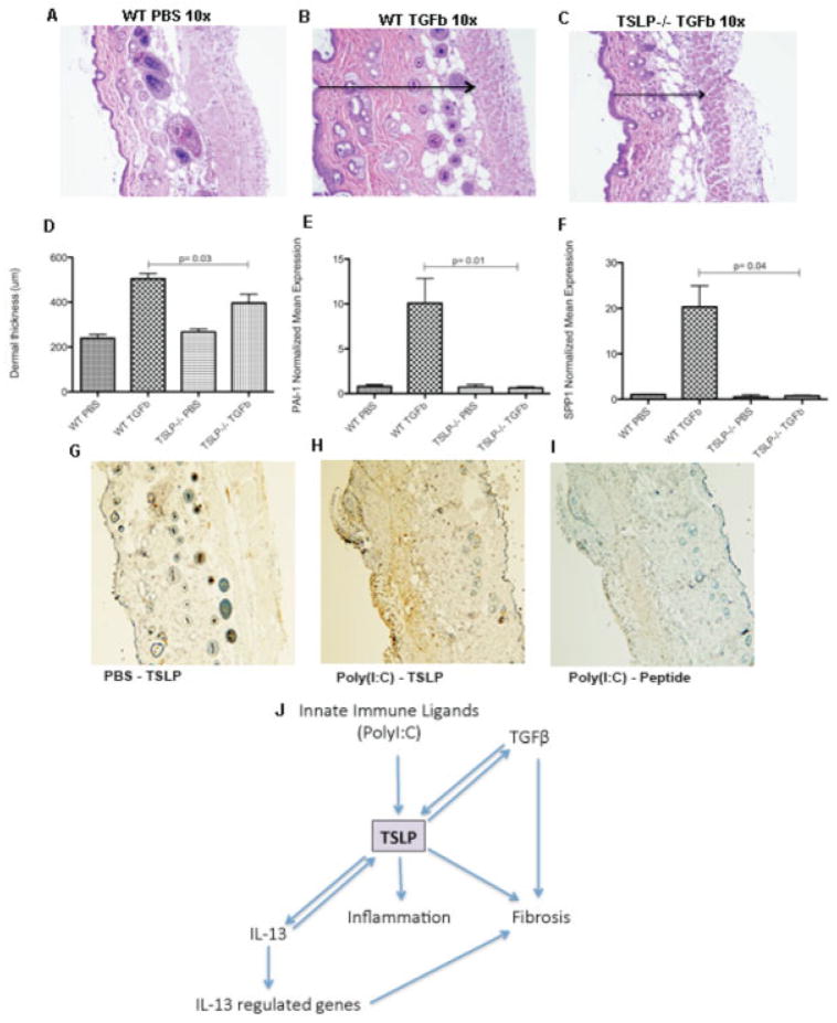
The in vivo profibrotic effects of transforming growth factor β (TGFβ) are partially dependent on thymic stromal lymphopoietin (TSLP). A–C, Representative images of hematoxylin and eosin–stained skin sections from a phosphate buffered saline (PBS)–treated wild-type (WT) mouse, showing preservation of the structures (A), a TGFβ-treated wild-type mouse, showing the dermal thickness (arrow), measured as the distance between the muscle and the epidermal layers (B), and a TGFβ-treated TSLP−/− mouse, showing a reduction in the dermal thickness (arrow) (C). Original magnification × 10. D, Dermal thickness, measured as the distance between the muscle and the epidermal layers. E and F, Expression of mRNA for plasminogen activator inhibitor 1 (PAI-1) (E) and osteopontin (SPP1) (F). G–I, Representative images of TSLP-stained skin sections from a PBS-treated control mouse showing low TSLP expression and preserved presence of the annexes (G), a mouse treated with poly(I-C) for 28 days (systemic sclerosis immune model), showing strong expression of TSLP staining in brown and thickening of the dermis with loss of the annexes (H), and a poly(I-C)–treated mouse with peptide-blocked anti-TSLP control negative staining (I). Original magnification × 10. J, Schematic representation of the complex interaction of TSLP with interleukin-13 (IL-13) and TGFβ.

