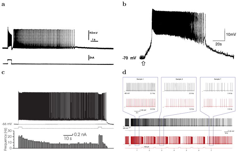
Figure 1.
Examples of slowly decaying firing rate in slice recordings. a. Cat layer 5 Sensorimotor Cortex after application of muscarine-a nonselective agonist of the muscarinic acetylcholine receptor (Schwindt et al., 1988). b. Acetylcholine-induced depolarization of Layer 5 pyramidal neurons of prefrontal cortex (Haj-Dahmane & Andrade, 1996). The arrow indicates the time of acetylcholine application. c. Layer 3 of lateral entorhinal cortex in the presence of muscarinic receptor activation (Tahvildari et al., 2007): membrane potential trace on the top, current stimulation trace in the middle and firing rate histogram on the bottom plot. d. Anterodorsal thalamus (area with a high percentage of head direction cells) after hyperpolarizing stimulus (current trace at the bottom), (Kulkarni et al., 2011).

