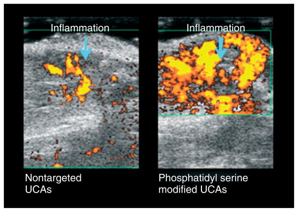
FIGURE 8.
Contrast-enhanced US images of a rat tumor obtained 7 days after performing the ablation therapy. Inflammatory region can readily be identified because of US contrast agent (UCA) targeting by phosphatidyl serine. The images were taken 12 min after injection of phosphatidyl serine UCAs. (Images courtesy of Dr. Agata Exner, Case Western Reserve University, Cleveland, OH, USA).

