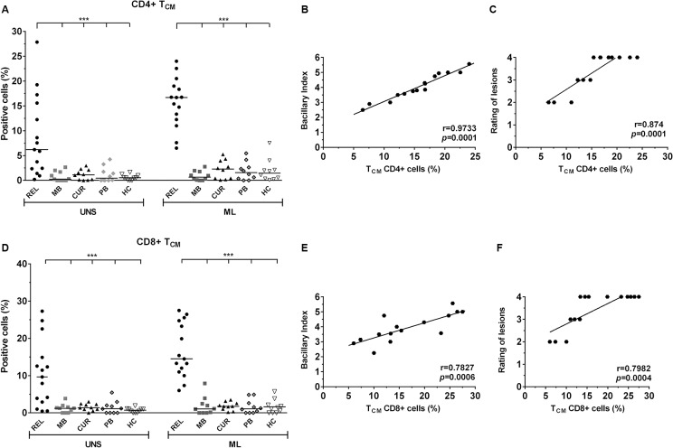
Fig 3. Increase in the frequency of CD4+ and CD8+ TCM lymphocytes in relapsed patients is related to BI and to the number of skin lesions.
CD4+ and CD8+ TCM cells (CD69+/CD45RO+/CD62L+high; A and D) in response to in vitro 24-h PBMC stimulation with M. leprae (20 μg/mL) by multiparametric flow cytometry (FACSAria, BD). The results are expressed as percentage of positive cells, and the statistical differences are shown (***p< 0.001; UNS: unstimulated). The horizontal bars represent the median of each group. In each analysis, isotype controls were used to distinguish between positive and negative cells. SEB (1μg/mL) was positive in all tests (data not shown). The Kruskal-Wallis test was used for comparison of stimulated cells with unstimulated cells, and the Mann–Whitney test to group comparisons. Correlation analysis between CD4+ and CD8+ TCM and BI (B and E) and the number of lesions (C and F). Significant positivity is demonstrated (r = correlation coefficient; p= significance level; Pearson test).

