
Fig. 2.
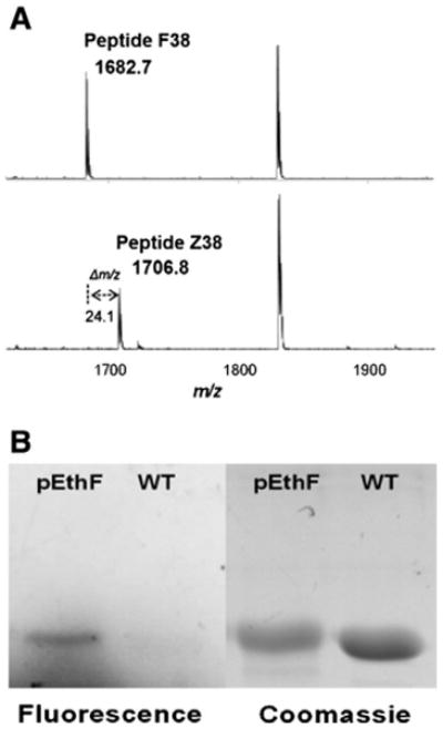
Site-specific incorporation of pEthF into the mDHFR-38Am and CuAAC-mediated coumarin-labeling. (A) MALDI-TOF analysis of trypsin-digested mDHFR-WT and mDHFR-pEthF. Peptide F38 (top) of the mDHFR-WT and Peptide Z38 of the mDHFR-pEthF (bottom). (B) Protein gel images of the fluorogenic dye-treated mDHFR-pEthF (pEthF) and mDHFR-WT (WT). The gel was subjected to UV (390 nm) irradiation to excite the fluorophore (fluorescence panel), and then stained with Coomassie brilliant blue (Coomassie panel) to visualize proteins.