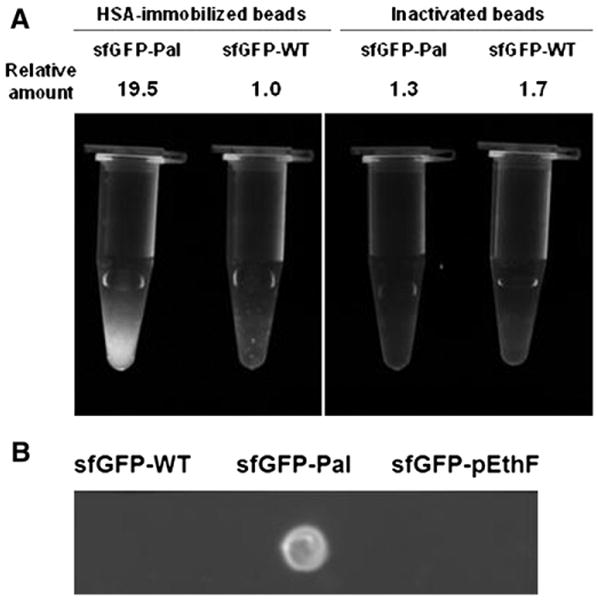
Fig. 4.

Relative albumin-binding affinities of sfGFP-variants. (A) Inactivated (amine-reactive functional groups blocked by glycine) or HSA-immobilized agarose beads were mixed with the sfGFP-WT and the sfGFP-Pal. After washing extensively with PBS, the fluorescence image was taken on the UV epi-illuminator at λex = 480 nm, and emitted light above 510 nm was captured. For a quantitative fluorescence measurement, the same amounts of agarose beads were loaded on a 96-well microplate and read on the plate reader at λex = 480 nm and λem = 510 nm. The relative amounts of sfGFP samples were calculated from the relative fluorescence intensities. (B) Four micrograms of each protein in 2 μL of PBS were dotted onto the HSA-coated nitrocellulose membrane and air-dried. After washing in PBS for 5 min and air-dry, the membrane was epi-illuminated at λex = 480 nm, and emitted light above 510 nm was captured.
