
Figure 1.
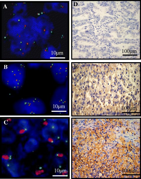
FISH analysis of the MET gene and immunohistochemical staining for MET protein. Gene copy numbers (GCNs) and amplification of the MET gene were examined by fluorescence in situ hybridization (FISH). (A) FISH negativity was defined as mean MET per cell < 5 copies. (B) High polysomy was defined as 5 copies≦mean MET per cell. (C) Amplification was defined as 2≦MET gene (red)/CEP7q (green) per cell. FISH positivity consisted of high polysomy and amplification. (D) Low level of MET protein expression in lung adenocarcinoma tissues. (E) Moderate level of MET protein expression in lung adenocarcinoma tissues. (F) High level of MET protein expression in lung adenocarcinoma tissues. Bars indicate 100 μm.